Analytics: Microscopy and The Royal Microscopical Society
Under the microscope: an interview with Peter O’Toole
IPT caught up with Peter O’Toole, president of the Royal Microscopical Society, to hear how his first few months as president have gone, his plans for the Society and how the field of microscopy might develop over the next few years
IPT: Before your presidency, you were the vice president for a number years at the Royal Microscopical Society (RMS). In what ways has your time as VP aided you in your new role over the past few months?
Peter O’Toole (PO): A big advantage of being able to shadow a president over that length of time is that you get to understand in depth what has been tried, what to try and what could be tried. It allows you to become familiar with the workings of the society at that level and how to use them to the best advantage. So then, once becoming president, it becomes easier to start making changes as you already know what is and isn’t possible.
In addition, a little bit of naivety and creativity are also helpful, as fresh ideas and new approaches are also important.
However, there’s a fine balance between simply being another iteration and introducing no changes or ideas, and being too much of a rookie and not understanding what problems could be encountered.
IPT: What part of your new role excites and inspires you the most?
PO: It’s exciting, but it’s also scary! The presidency is a big task and the previous presidents have their reputations and were very positively influential for the society.
The biggest concern is not to break what isn’t broken, and I’m very cautious of balancing that with my ambitions for the society. But it is exciting that each of the previous presidents have all thought differently and approached the presidency in their own ways. They all had different ways of working, and they all had subtly different ways that they wanted the society to evolve. That’s what is so good about changing presidency, as the presidency comes in and will evolve one or two aspects, allowing the society to grow and flourish. The presidencies always enable different evolutions within the stability of the executive committee, while ensuring that new ideas are good for the society. It is the responsibility of the president to grow the society and help it evolve, and this ability is what excites me the most about my presidency.

“ AI being used for analysis and prediction is going to have the biggest impact going forward. It’s probably going to be the most influential change that we’ve seen for some time ”
IPT: What are your plans and visions for the RMS as president?
PO: Firstly, one goal is to try and grow the society’s membership. We have quite a large membership, yet there is a significant number of people that attend our courses, events and conferences who aren't members, but for whom the RMS is still important. It would be great if those people become full members – their support from additional membership fees would sponsor and enable even more of those activities, hopefully drawing even more people into the RMS.
Similarly, the society is fortunate to have an active volunteer base and we'd like to engage with the volunteers to a greater degree than we currently are. This would allow us to deliver more back to the community, as well as support those who do volunteer. We can offer help and guidance to volunteers in their professional careers and they can help the Society within the microscopy world – a win-win! Embracing more volunteers, developing new outreach and introducing more courses and events are all things that we’d like to see develop over the next three to five years.
The second goal has to do with how the RMS was started and where the world is today. When the RMS was started, it became very popular very fast, especially at an international level, as it was the only society of its nature covering these topics. We had people from all over the world joining, who all have been and still are key members of the society. As the microscopy field has grown, people have started more local alternatives to the RMS, yet it still remains the largest international society. We have a lot of international members, but we don’t often deliver enough events and offerings outside the UK (for a variety of good historic reasons). Now, the world is getting smaller again, especially with virtual events. During the COVID-19 pandemic, we organised meetings in over 35 different countries with over 1,000 different scientists, including not only RMS members but also microscopists across the world. The ideal would be to capitalise on this and grow a bigger international movement, facilitate wider involvement and support different players. In these meetings, the RMS helped run them alongside the academic leads from the host country; I’d like to see a continuation of this now that the pandemic has wound down, as well as have the RMS be more active and offer more support on the international level.
IPT: You head the Imaging and Cytometry Labs at the University of York and your students could be the future leaders in the field – do you feel that they engage with microscopy and cytometry in the same way you did when you were studying?
PO: The amount of time that I spend face-to-face with PhD students is now much less than it was, as my team is extremely expert and takes on a lot of that responsibility. However, I can definitely say that some of the people who have come through that leave as great scientists. They’re fantastic microscopists within that subset and actually their career may end up having more to do with the technology itself than the scientific questions they would be investigating. This has definitely been the case over the last few years and we’re still seeing those type of people come in and go through platforms – possibly, even increasingly so. We now have more microscopes in our labs than ever before, in quantity and variety, giving us more capacity for teaching. There are also more frequent moves to embrace new technologies and approaches to data analysis and microscopy – which, in addition to being admirable coming from the students, is a credit to my team for inspiring them to do that, as well as keeping them at the forefront of the technology.

Some of these PhD technologists are some of the best because they’re not just based on microscopy or cytometry; they’re going on to do the single cell transcriptomics or to do spatial lipidomics. They’re bridging research gaps and conducting all the other correlating techniques and other technologies that are out there. It’s really good to see how students are able to pick up all those different technology platforms and to run their samples from start to end, to all those different varieties of labs – genomics, the tableau, proteomics, the imaging and the cytometry – and it’s quite some skill. For me, part of the enjoyment of being a scientist is the ability to exploit technology in ways that enable you to answer questions that couldn’t otherwise be answered, in the best way. This is definitely something we see in these students, and those that are multifaceted with the technologies receive some of the biggest impacts and best results.
IPT: What do you find most rewarding when teaching the RMS Flow Cytometry and Light Microscopy courses?
PO: It’s having dedicated time to just engage with students again. The students that come on the RMS Flow Cytometry and Light Microscopy courses are not just PhD students – they’re technical staff, postdocs and academic staff – there’s quite a broad spectrum. I like the fact that the fundamentals of the courses don’t change, and that we welcome both complete beginners and experienced users. So many of us get launched into it at some level, which can mean an understanding of the basics is missing, as you’re thrown straight into it and have to learn while doing. It’s quite nice to be able to reassure the students that they know what they’re doing and let them understand why it’s being done like that. Moments like that are quite nice, as are those when you show them what more their microscopes and cytometers can do. It’s really empowering to know that they’ll go back with new approaches to their research. Part of the joy comes from those interactions with people and so, at the end, it’s quite sad once everyone leaves because you’re with each other so much over the course.
IPT: The Imaging and Cytometry Laboratory at York, UK, is seen as one of the top European centres and the lab has the very latest equipment. Over the years, what has become your favourite piece to work with and why?
PO: With flow cytometry, in the early days, the MoFlo Cell Sort and the Lasik System was a difficult beast at times, but once you really became at one with it, it was like a pet. That was a really lovely time as it wasn’t so hectic at work, and so that was one of my favourite instruments. The other must be the LSCM 510, even though the technology has moved on and it wouldn’t be able to compete today. But again, it was a real workhorse and we spent so much time on it. I could’ve challenged anyone to get an image out of it more quickly and we knew how to run it with our eyes closed. But those days have passed and the newer technologies are far better than they were, but if I had to pick favourites, I’d look back to those two.
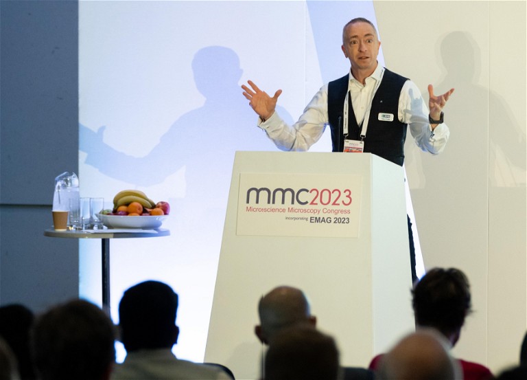
IPT: How has the RMS Microscience Microscopy Congress (MMC) changed since its inception in 2014?
PO: Going back even further than 2014, Microscience was an event itself that used to be held in London, and it was mostly an exhibition with a few lectures and seminars. The scientific programme became stronger and stronger, which helped to boost the exhibition as well – to go to just an exhibition is a bit of a luxury, so what better way to get people in than to link it with a scientific conference. The exhibition is still free, so anyone can just turn up to the exhibition and have a look around, but they could, if they wanted to, then go to the science side of it as well.
It’s been really nice to see the incorporation of the Electron and Microscopy Group (EMAG). They meet every year, but every other year, it’s held within the MMC. It’s an example of what I love about MMC – it’s become an umbrella of different events. This year had the largest number of delegates present at an MMC, which was great – it’s going in the right direction! The quality of the science on display and the exhibition hall were both excellent.
It’s always evolving as well, which is amazing to see. It might look similar every year but, behind the scenes, it’s always evolving: the ratios, the proportions and what’s delivered. It’s important not to get too niche sometimes – there are quite a lot of people who think that their specific focus is what everyone’s interested in, and the MMC Organising Committee has to ensure the topics and content appeal to a broad audience while ensuring that those niche markets can also be addressed. On the whole, the event has taken quite a step forward with growth this year, so we’re all very excited for the next one in 2025.
IPT: What impact do you see artificial intelligence (AI) having in the future of microscopy?
PO: AI being used for analysis and prediction is going to have the biggest impact going forward. It’s probably going to be the most influential change that we’ve seen for some time. It’s still very much in its infancy, but it is maturing incredibly fast, which is both exciting and daunting at the same time.
I don’t think it’s going to mean we'll be out of a job, so that’s good! People in the imaging world are developing machine learning algorithms and looking at AI to help us as users. This means that downstream analysis can be more powerful, faster and more accurate, which should allow users to tease out information that would not otherwise be visible.
IPT: What challenges do you see facing microscopy over the next few years and how can they be overcome?
PO: One of the frustrations of the last five years has been the pace of AI development and adoption. It’s been good in some areas, but everyone wants something different, and so one of the biggest challenges is to find or create solutions for everyone. And that’s not trivial.
Additionally, even though the community is fairly big, the user base is not on the same scale as it could be, and I think the speed and volume of throughputs is going to be critical to open up those avenues. Similarly, the diversification of techniques – the correlating microscopy to spatial transcriptomics, spatial lipidomics, etc – and their embracement can be quite beneficial. These are big steps and big challenges but progress has already started, so it’ll be interesting to see where that is in five to seven years; it’ll look very different to how it is today.
Peter O’Toole is the president of the Royal Microscopical Society (RMS) and leads the Imaging and Cytometry Labs at the University of York, UK.